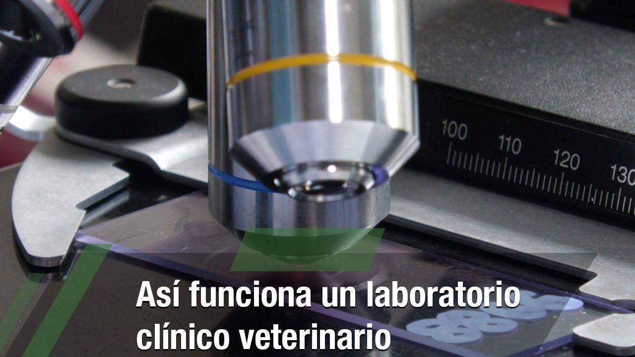
 En Southeast Veterinary Neurology, con cierta frecuencia revisamos tomografías computarizadas y resonancias magnéticas efectuadas en otros centros que brindaron un diagnóstico incorrecto. En segundo lugar, la mayoría de los centros humanos efectúan 30 o más resonancias magnéticas al día. Segundo, la mayoría de las instalaciones para humanos son reembolsadas por las aseguradoras. Primeramente, la mayoría de las instalaciones para humanos son reembolsadas por las empresas aseguradoras. 648 estructuras anatómicas identificadas en estas imágenes radiológicas por laboratório clíNico veterinário el Dr. Antoine Micheau, distintas colores para hacer más simple la lectura y la búsqueda de construcciones anatómicas en cada radiografía. Nuestro hospital está autorizado para llevar a cabo las radiografías por el CEPA, por lo que si lo necesitas se tienen la posibilidad de efectuar sin problema. Los rayos X penetran el cuerpo del perro y de las personas que participan en la prueba, dañando las células irradiadas.
En contraste a la radio, en la ecografía no se usan rayos X, sino más bien ultrasonidos. Deja el examen de los órganos en movimiento, no estáticos, y no piensa riesgo de radiación para el perro. Algunas consultas veterinarias están equipadas con la última tecnología, lo que enseña por qué los costes son más altos que en otras consultas. Es muy útil en casos de urgencia y a veces es requisito la utilización de medios de contraste como en problemas meaderos o gastrointestinales, primordialmente. El TAC se basa en los principios de la radiología, generando rayos X, con lo que produce radiación ionizante, por lo que es primordial la radio protección de la salón. Una radiografía del esqueleto requiere casi siempre regentar tranquilizantes al perro o gato, ya que es esencial que el animal se sostenga inmóvil durante el trámite.
Por qué podrías necesitar considerar un seguro para mascotas
No obstante, el incremento de mA suele provocar una mayor carga térmica en los tubos de rayos X, lo que limita los tiempos de exposición y reduce la vida útil del tubo, además de aumentar la exposición del paciente a la radiación. Tanto para los pacientes con gatos para los controles, se recolectaron muestras de resección de colon de espesor terminado, fijadas en paraformaldehído al diez% y luego incluidas en cera de parafina. Se consiguieron partes de 5 μm y se procesaron para una tinción estándar de hematoxilina y eosina como se describió anteriormente (14). En exactamente las mismas secciones teñidas, se midió el grosor de la mucosa muscular lisa y de ámbas capas de musculatura externa lisa (capas musculares circulares internas y longitudinales externas) utilizando el software Image J (NIH, Bethesda, Maryland, EE. UU.).
Atlas de interpretación radiológica en pequeños animales
Con las películas de grano grande y las pantallas de cristal grande ocurre lo opuesto. Los tres parámetros anteriores son interdependientes, y el tiempo de exposición y los mA son tan esenciales que se frecuenta usar el término miliamperios-segundo (mAs) para indicar el producto de estos 2 componentes. Aumentando los mA y reduciendo el tiempo de exposición en una cantidad proporcional, se consigue una radiografía con menor posibilidad de degradadarse por el movimiento. Por regla, es mejor minimizar el tiempo de exposición pero mantener unos mAs y una escala de contraste apropiados. Al aumentar el kVp se incrementa el número de fotones que penetran en el sujeto y, por consiguiente, se oscurece la imagen. La radiología veterinaria es una técnica de diagnóstico por imagen muy utilizada por ser un trámite sencillo de efectuar, comunicando velozmente sobre el estado de tejidos blandos, huesos o articulaciones.
Because of the necessity to remain still for a relatively very lengthy time while scanning is accomplished, animals undergoing a CT scan are anesthetized. Radiology is a branch of drugs that uses imaging to diagnose and deal with illness. The following websites may help you better perceive the numerous exams that fall underneath the Radiology heading. In the Dominican court system, after the prosecutors file charges, a judge holds a listening to to find out if the evidence warrants the case advancing to a trial or being dismissed, a course of that would take several months. Franco was charged last month with sexual abuse, sexual exploitation of a minor and human trafficking, stemming from a relationship with a then-14-year-old lady that began in December 2022. [newline]Franco, who was 21 on the time, might withstand 20 years in jail if convicted. But when you can keep them away from your dog (and everyone else in your family), you’re higher off. If your canine has only delicate signs after ingesting the insect, don’t assume you’re in the clear simply but.
Choice of the right pace system for a selected use is based not only on the world being radiographed but in addition on the capabilities of the machine. Small, transportable x-ray machines can be utilized for larger physique elements with quick film-screen combos, substantially bettering the utility of these machines. Higher kV settings produce more penetrating beams during which a better share of the x-rays produced penetrate the subject being radiographed. There can be a decrease in the proportion difference in absorption between tissue sorts. This results in decreased distinction (long-scale contrast) on the ultimate image. High kVp techniques are most helpful for studies of physique regions with many different tissue densities (eg, thorax).
PREVENT YOUR DOG FROM TROUBLE WITH BEES AND OTHER STINGING INSECTS
Because images stored in a digital format are easily manipulated by varied pc packages, it is possible that they could probably be altered (accidentally or deliberately) to replicate a unique situation than the actual one. For this purpose, many digital image formats usually are not acknowledged as legal documents and are not acceptable in a court docket of legislation. DR systems have been developed that don't require a cable to communicate between the detector and the computer processing the data into a picture. The cable has been changed by wi-fi communication on specified electrical magnetic frequencies which would possibly be unlikely to be interfered with by other electromagnetic gadgets corresponding to cell telephones and digital tools.
The Purpose of a Technique Chart for Veterinary Radiography
The tube present, measured in milliamperes (mA), and voltage, measured in kilovolts (kV), decide the strength and number of x-rays produced and are two of the three exposure elements that might be set on most x-ray machines. Kilovoltage potential (kVp) is the highest potential voltage achieved at any given kV setting. The aim of canine radiographs is to determine a analysis, or get hold of a final answer with out having to perform further, extra invasive checks or procedures. Just like MRI tools, CT scan tools may be very expensive, massive and requires skilled technicians to operate. However, and in our mission to verify all diagnostic imaging instruments can be found for your loved one canine companion to profit from, we provide CT scans for canines at our veterinary hospital. For the procedure, the animal is placed in a tubular electromagnetic chamber and pulsed with radio waves, causing tissues in the physique to emit radio frequency waves that might be detected.
Radiography in veterinary practice- a review and update by Kimberly Palgrave
The emitted waves are then transformed into photographs which would possibly be displayed on a computer display screen. Sequential examination of slices by way of the physique is done in the identical method as for computed tomography. Because the process is quite prolonged and the animal must not transfer all through the process, general anesthesia is used generally. In this process, the animal is placed on a motorized bed inside a CT scanner, which takes a series of x-rays from different angles.
How can I prepare my dog for their X-ray appointment?
Monitoring of exposure also offers evidence of correct adherence to radiation safety requirements if questions come up as as to if an employee’s medical condition might be associated to radiation exposure. Proper positioning is also important to maximize the diagnostic content material of the x-ray examination. In many cases, improper positioning or radiographic examination may end up in a misdiagnosis or lack of ability to understand major lesions. Both right and left lateral recumbent radiographs are beneficial in dogs and cats. This is completed as a result of positioning of the animal on its aspect leads to rapid relocation of fluids to and atelectasis of the draw back lung.