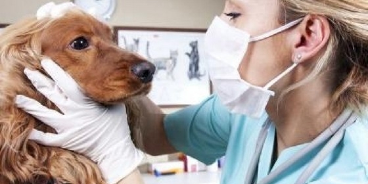
¿Cuál es el costo de una ecografía para el embarazo de perros?
Entre las primordiales ventajas de la ecografía es que es un trámite seguro y sin efectos secundarios conocidos. En contraste a otras pruebas de diagnóstico que comprometen radiación, como los rayos X, las ecografías no exponen a su perro a radiación probablemente dañina. Esto lo transforma en una alternativa preferida, especialmente para perras preñadas o aquellas con problemas médicos preexistentes. Si bien el costo de una ecografía para un perro puede ser una inversión importante, es importante priorizar la salud y el confort de su mascota. La detección y el diagnóstico tempranos de dolencias médicas pueden conducir a un tratamiento más eficaz y mejores resultados para su amigo peludo. El precio de una ecografía cardiaca tiende a ser más prominente gracias a la dificultad del procedimiento y el equipo especializado necesario, pudiendo oscilar entre los cien y los 300 euros.
 The 3 Principles of Radiation Safety
 Incompletely formed tracheal rings and a flaccid or redundant tracheal membrane cause narrowing of the lumen. This is usually a dynamic lesion that is it changes with the part of respiration. On inspiration there is negative stress throughout the cervical portion of the trachea and it'll collapse or narrow. On expiration, positive strain within the thorax causes narrowing or collapse of the intrathoracic trachea and in some circumstances the primary stem bronchi. Uniform narrowing of the tracheal lumen is seen in tracheal hypoplasia, particularly in brachycephalic breeds. Apparent uniform narrowing of the trachea may be seen in canines due to hemorrhage attributable to intoxication by vitamin K antagonist rodenticides. In these circumstances, the air shadow of the lumen might be a lot smaller than the outline of the tracheal rings.
The main pulmonary artery is not seen usually as a separate structure, however when it dilates sufficiently in dogs, it will appear as a focal bulge within the 1 o’clock place in VD or DV views (Fig. 32-13).
Fat is noted on this ventrodorsal radiograph of a small-breed dog between the right cranial and right middle lung lobes (white arrows). Note that the fat within the fissure is thickest centrally and thinnest peripherally. This appearance is the other of that of pleural effusion, by which the widest portion of the pleural fluid is peripheral and thins because it extends medially within the pleural fissure line. Right lateral (A) and ventrodorsal (B) pictures from a dog with generalized megaesophagus secondary to myasthenia gravis. Note the ventral deviation of the trachea and coronary heart in the cranial and middle mediastinum. In addition, the dorsal, ventral, and lateral borders of the caudal thoracic radiograph converge to the esophageal hiatus within the central, laboratórios farmacêuticos VeterináRios dorsal third of the diaphragm (arrows). In addition, a 3-view thorax (right lateral, left lateral, and dorsoventral or ventrodorsal view) is considered the standard of care in veterinary drugs.
Thoracic Radiographic Exposure
The Customer will be succesful of entry an invoice in digital format on his account inside the platform. It is really helpful to keep the order affirmation web page, to find a way to maintain the elements composing the order, in addition to the order number. The Licensee acknowledges that the granted use solely permits the Software to be consulted on the Website or throughout the Applications downloaded onto the mobile system (tablet, telephone) belonging to the Licensee. The Licensee agrees that this License does not embody any additional providers aside from the Paid Services "outlined" in Article 1.1 "Definitions". The objective of this contract is thus to determine the situations of subscription by the Customer permitting him to make use of the Paid Services of the Products, accessible from the Site or the Applications, throughout the limits of use defined in the License.
What Does a Chest X-ray Reveal in Dogs?
Like different veterinary services, X-rays are usually dearer in urban and extremely populated locations than in rural locations. And the worth of X-rays can differ by the veterinary practice in the identical city, so you could benefit from buying round. Some canines in excessive ache or are feeling nervous won’t have the flexibility to sit nonetheless sufficient to get a clear X-ray. If this is the case together with your pup, sedation or anesthesia could additionally be required. Other causes for sedation embrace if the dog’s muscle tissue need to be relaxed to get a transparent image or when the X-ray is of the spine, skull, or tooth. The further price for sedation or anesthesia can vary from $40 to $200. Poor penetration of the spine, particularly superimposed on the scapula, ought to warn you to this variation.
Then, a call has to be made if the pulmonary parenchyma is just too opaque or too radiolucent. Often, a mix of pulmonary patterns is current, and in those instances it is most efficacious to find out the predominant sample as it'll greatest define the supply of the issue. Horizontal beam views are helpful for a variety of thoracic issues. The affected person could additionally be positioned in lateral recumbency, and a VD view utilizing a horizontal X-ray beam taken. This view permits redistribution of fluid and air, aiding in the diagnosis of delicate pneumothorax or pleural effusion. Evaluation of suspected chest wall or mediastinal lots could also be enhanced when pleural fluid falls away from the world of interest. An different position with the patient held upright, or "sitting", may be done using a VD view and horizontal X-ray beam.
Filmless Radiography for Diagnostic Imaging in Animals
They are also indicated in geriatric patients, and in sufferers that will have cancer, to evaluate for metastasis (spread). X-rays of the chest must be taken of each animal that has been hit by a automobile or suffered other kinds of major trauma as a end result of they can reveal many types of accidents to the chest wall, Joao-Vicente-Soares.Blogbright.net lungs and heart, or other injuries like diaphragmatic hernia. X-rays are additionally typically repeated to observe progress after treatment or after eradicating fluid for higher visualization of constructions. Even normal outcomes assist determine well being or exclude certain diseases. Based on the place of the heart, the place is the apex situated on the lateral and the VD/DV radiograph? The apex is one of the best exterior cardiac landmark that documents the division between the left and proper sides of the cardiac silhouette. Remember although that the exterior structure that one is taking a look at is really the outer border of the pericardial lining and not the epicardial surface of the guts.
Additional Views
But they can not definitively diagnose harm to the cruciate ligament, the meniscus, or both. Other risks, although minimal, can embrace errors in interpretating the results and misdiagnosis, particularly with subtle modifications. Therefore, it’s always greatest to have the X-rays reviewed by a board-certified radiologist. X-rays, put merely, are 2D photographs of a 3D object, a sort of electromagnetic radiation produced when electrical power from electrons is converted into X-rays inside a specially designed tube. Neither sedation nor anesthesia is required in most patients; nonetheless, some pets resent positioning for an X-ray and might have tranquilization or ultrashort anesthesia. In a couple of states there's a authorized requirement for sedation in order that personnel aren't uncovered to any X-rays while holding an animal patient. However, in most cases, the unsedated pet is attended by assistants who put on acceptable lead-shields to reduce their exposure to X-rays.


